Hamamatsu Photonics K.K. (Hamamatsu City, Japan) has developed a new instrument, creating a unique class of cell analyzers. By implementing light-sheet optics and image analysis, in combination with an easy-to-use interface, we have engineered a fast, user-friendly system ideal for 3D cell screening assays.
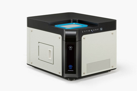
Imaging is a critical part of scientific research, from benchtop discovery to preclinical drug screening1. Building on our deep foundation of engineering and life science applications, we have developed a new tool for advanced live cell image analysis. The CYTOQUBE Light-Sheet Microplate Cytometer (C15200-01RGBU) is designed to enable fast, information-dense imaging of multi-well cell culture plates2,3. Using a streamlined implementation of light-sheet microscopy and our own Zyncscan image processing platform, the CYTOQUBE rapidly acquires 3D fluorescence images of each well in an entire microplate4. The imaging data collected with the CYTOQUBE and Zyncscan enables rapid assays for size, shape, number, and color/intensity of cells or cell populations. The CYTOQUBE's advantages start with the light-sheet optical system for simplified 3D acquisition and culminate with our advanced Zyncscan imaging processing algorithms for fast acquisition and rendering of 3D images and increased signal-to-noise ratio through the removal of background fluorescence.
Since the CYTOQUBE uniquely excels at fast 3D imaging of microplates, this may make it especially useful in drug discovery. The full path from target identification to drug development can take 12-15 years and is both very high risk and high reward. Classic 2D cell culture drug screening assays are well-established and achieve high throughput, but notably lack in vivo relevance. To enhance successful outcomes, especially for cancer drug development, scientists are increasingly turning to 3D cultures of spheroids or even organoids. While these cultures more closely model in vivo conditions, they are also more difficult to assay especially because 3D imaging adds significant time to results. Any tools or processes that can speed up the acquisition of live 3D cytology data could add significant benefits in the drug discovery pathway. Because the CYTOQUBE is able to capture fast 3D fluorescence image data, it has the potential to boost the efficiency of drug screening and opens up new possibilities for cellular assays.
Principle of CYTOQUBE and Zyncscan Technology:
Cells in the microplate are illuminated from directly below with light-sheet excitation, and XZ tomographic images are captured by a scientific CMOS camera. The detection is decoupled from the illumination and situated at an oblique angle to the sample and the illumination. By continuously acquiring XZ tomographic images while moving the microplate in a direction perpendicular to the light-sheet illumination, a full 3D fluorescence image is constructed in one scan of the plate. In addition, the CYTOQUBE software is able to remove, in real time, unwanted background fluorescence that can degrade the signal, while simultaneously acquiring XZ tomographic images.
For more information please visit https://www.hamamatsu.com.